
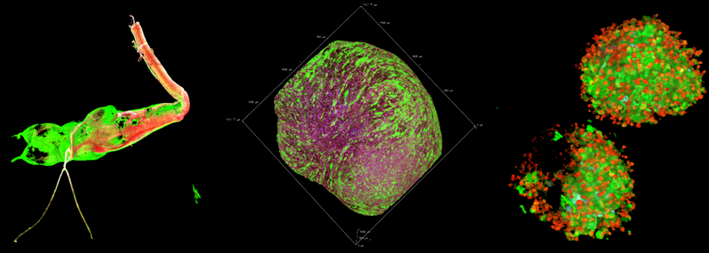
Imaging of living cells is mainly carried out by fluorescence microscopy. Wide field microscopy, which allows flexible excitation and rapid acquisition.
Technics are generally used to visualise the dynamics and development of cells over long periods of time. Confocal microscopy will allow the study of subcellular dynamic events.
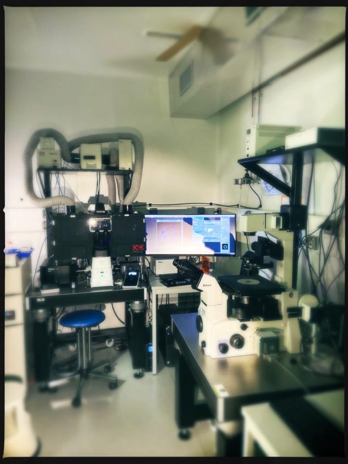
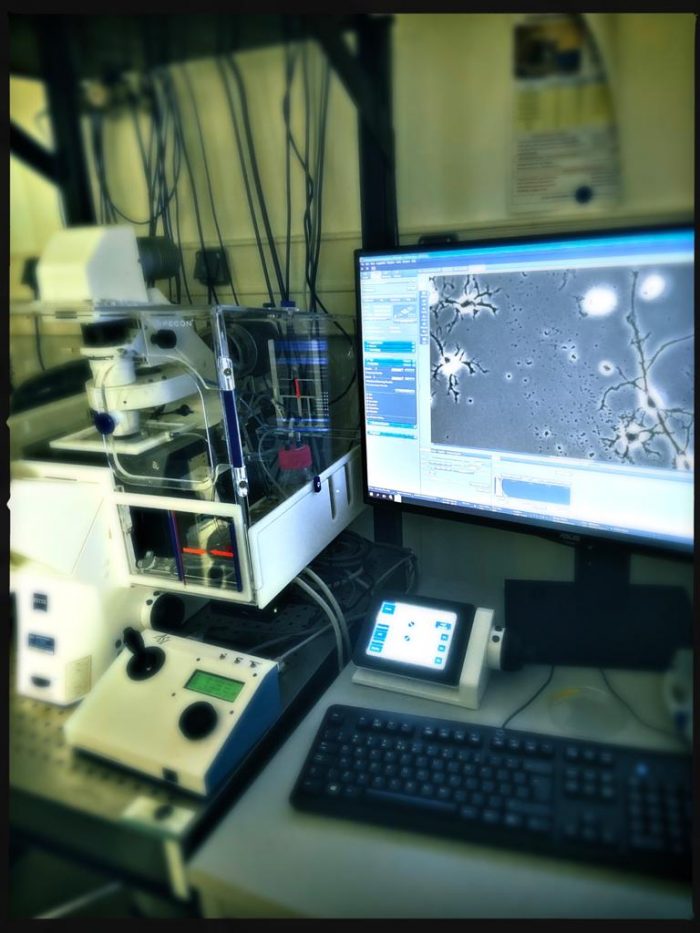
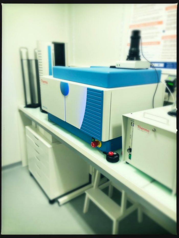
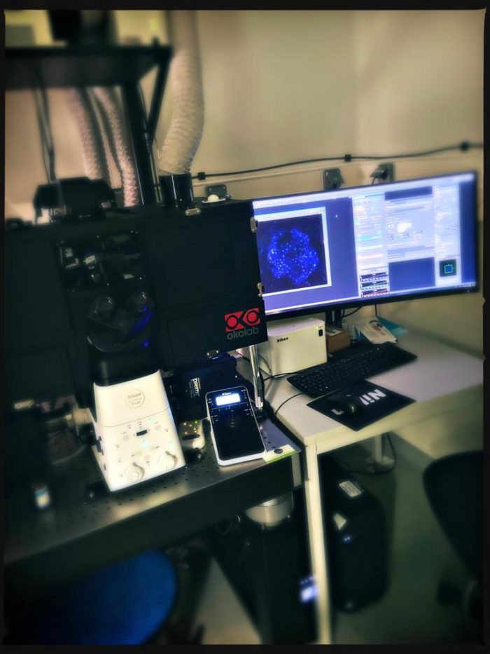
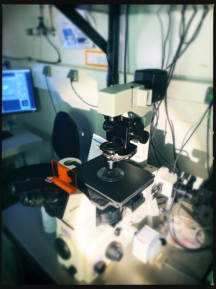
The CELIS core facility of the Brain Institute has all the equipment and techniques required to increase the efficiency of your living cell imaging process with inverted microscopes for cell and tissue cultures.
Guidance for equipment selection.
Training on equipment for stand-alone use.
Scientific/technical assistance.
Development of personalized protocols for image acquisition/analysis.
Assistance with result interpretation and protocol writing for imaging.
Interface with suppliers of equipement when is necessary
 |
 |
 |
 |
 |
 |